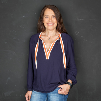
SPEAKER PROFILE |
 Prof. Laura Suter-Dick School of Life Sciences, FHNW Switzerland |
Electrospun extracellular matrix scaffolds for neural regeneration
Abstract
Decellularization of tissues, with the goal of applying extracellular matrices (ECM) based scaffolds in regenerative medicine, requires the preservation of the complex biochemical cues specific to the native microenvironment (1-4).
In this work, cell-free, spinal cord-derived ECM scaffolds were produced to support the alignment of neural progenitor cells and neurons. Decellularization of pig spinal cord was accomplished by a combination of mechanical, physical (freeze-thawing) and chemical (SDS incubation) treatments. The decellularized tissue was characterised via dsDNA quantification and histological staining to assess the presence of cell nuclei, proteins and glycosaminoglycans. Successfully decellularized spinal cord was lyophilized, milled and solubilised into a bioink, using hexafluoroisopropanol (5 % w/v ECM + 0.1 % w/v Polyethyleneglycol) and patterned into an electrospun fiber scaffold (using a 3D Bioprinter). The results show that porcine spinal cord can be successfully decellularized leading to an ECM with less than 50 ng/mg dsDNA, and without cell nuclei. The formulated bioink enabled spinning of acellular spinal cord ECM scaffold with fibers of <10 µm diameter. The non-cytotoxic scaffolds supported the viability of a human neural cell line (SH-SY5Y) over the course of 14 days and the selective differentiation into neurons, as confirmed by immunolabeling of specific cell markers (HB9, Tubulin ß). The combination of biochemical and topographical cues in this patterned ECM-derived scaffold induced the alignment of neural cells in vitro.
The strategy of combining decellularized tissues followed by specific deposition patterns to optimize biochemical and topographical cues opens the way for clinically relevant central nervous system scaffolding solutions.
----------
References
(1) Crapo et al. Biologic scaffolds composed of central nervous system extracellular matrix, Biomaterials, 2012.
(2) Keane et al. Methods of tissue decellularization used for preparation of biologic scaffolds and in vivo relevance, Methods, 2015.
(3) Volpato et al. Using extracellular matrix for regenerative medicine in the spinal cord, Biomaterials. 2013.
(4) Badylak et al. Extracellular matrix as a biological scaffold material: Structure and function, Acta Biomaterialia, 2009.
During her career, she specialized in advanced in vitro systems for toxicity assessment, alternatives to animal methods, and toxicogenomics. Recently her work focused on tissue engineering and the application of microfluidics and microphysiological systems. She is actively involved in teaching and supervises bachelor, master, and Ph.D. students. She is president of biotechnet Switzerland, and a member of the Swiss Centre for Applied Human Toxicology (SCAHT) and the Swiss 3R Competence Center (3RCC). She is vice president of the Swiss Toxicology Society and member of ESTIV.
In this work, cell-free, spinal cord-derived ECM scaffolds were produced to support the alignment of neural progenitor cells and neurons. Decellularization of pig spinal cord was accomplished by a combination of mechanical, physical (freeze-thawing) and chemical (SDS incubation) treatments. The decellularized tissue was characterised via dsDNA quantification and histological staining to assess the presence of cell nuclei, proteins and glycosaminoglycans. Successfully decellularized spinal cord was lyophilized, milled and solubilised into a bioink, using hexafluoroisopropanol (5 % w/v ECM + 0.1 % w/v Polyethyleneglycol) and patterned into an electrospun fiber scaffold (using a 3D Bioprinter). The results show that porcine spinal cord can be successfully decellularized leading to an ECM with less than 50 ng/mg dsDNA, and without cell nuclei. The formulated bioink enabled spinning of acellular spinal cord ECM scaffold with fibers of <10 µm diameter. The non-cytotoxic scaffolds supported the viability of a human neural cell line (SH-SY5Y) over the course of 14 days and the selective differentiation into neurons, as confirmed by immunolabeling of specific cell markers (HB9, Tubulin ß). The combination of biochemical and topographical cues in this patterned ECM-derived scaffold induced the alignment of neural cells in vitro.
The strategy of combining decellularized tissues followed by specific deposition patterns to optimize biochemical and topographical cues opens the way for clinically relevant central nervous system scaffolding solutions.
----------
References
(1) Crapo et al. Biologic scaffolds composed of central nervous system extracellular matrix, Biomaterials, 2012.
(2) Keane et al. Methods of tissue decellularization used for preparation of biologic scaffolds and in vivo relevance, Methods, 2015.
(3) Volpato et al. Using extracellular matrix for regenerative medicine in the spinal cord, Biomaterials. 2013.
(4) Badylak et al. Extracellular matrix as a biological scaffold material: Structure and function, Acta Biomaterialia, 2009.
Bio
Prof. Dr. Laura Suter-Dick is currently Professor for Cell Biology and Molecular Toxicology at the School of Life Sciences, University of Applied Sciences Northwestern Switzerland (FHNW). She is a European Registered Toxicologist (ERT), holds a Ph.D. in biology, and acquired more than 15 years of research experience in Industry before moving to academia in 2012.During her career, she specialized in advanced in vitro systems for toxicity assessment, alternatives to animal methods, and toxicogenomics. Recently her work focused on tissue engineering and the application of microfluidics and microphysiological systems. She is actively involved in teaching and supervises bachelor, master, and Ph.D. students. She is president of biotechnet Switzerland, and a member of the Swiss Centre for Applied Human Toxicology (SCAHT) and the Swiss 3R Competence Center (3RCC). She is vice president of the Swiss Toxicology Society and member of ESTIV.